

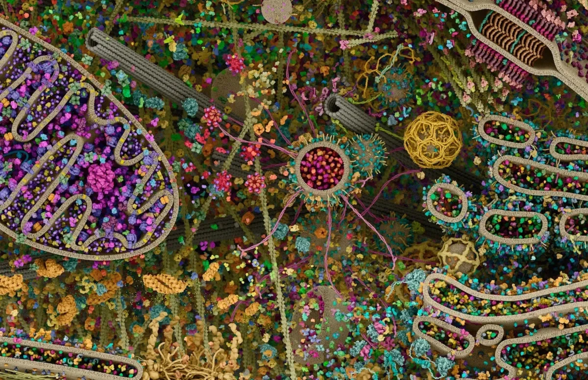
A social media post by the account Pubity, captioned "The most detailed look inside a human cell you've ever seen," had passed 173,000 likes when we captured it. It shows a glowing, cutaway sphere packed with thousands of colorful molecular shapes. It looks like a photograph, but it is not one. Visuals like this are computer renderings, and the tech behind them is a story in itself.
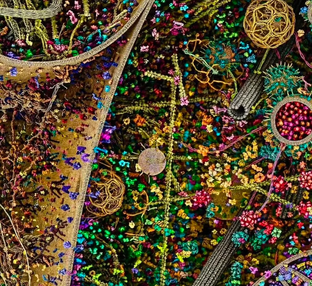
A rendering, not a photo
The visible text of the post does not name a creator, so we cannot trace this exact image to a specific team. The best-documented example of this genre is a cell landscape by scientific animators Gaël McGill and Evan Ingersoll of Digizyme. Newsweek fact-checked a viral version of it in 2022 and rated the "most detailed image of a human cell" claim false. The outlet described it as a 3D computer illustration of a eukaryotic cell, not a photograph.
McGill told Newsweek that a real photograph at that level of detail is not possible, because molecules at that scale are smaller than the wavelength of visible light. He also said the creators never made the "most detailed" claim. According to him, it was added after the image was taken from their portfolio.
Technical architecture: where the data comes from
According to My Modern Met, the Digizyme render was developed between 2009 and 2015. McGill describes it as modeled from X-ray, nuclear magnetic resonance (NMR) and cryo-electron microscopy datasets for its molecular components. In other words, the shapes of the individual proteins come from real lab measurements. Software then arranges them into a plausible cellular scene.
That also means the render was not made with modern generative AI. The human work is in the data and the design choices.
Where AI actually comes in
Machine learning does play a real role in cell visualization, just not in this particular image. In 2018 the Allen Institute for Cell Science launched the Allen Integrated Cell, which it called the first predictive and comprehensive 3D model of a live human cell. It combines two models:
- A probabilistic model that predicts the most likely shape and location of cell structures based only on the shape of the plasma membrane and the nucleus.
- A label-free model that predicts where 3D structures sit in cells without fluorescent labels. It was trained on 3D images of thousands of gene-edited cells carrying fluorescent protein tags, and it can work on brightfield microscopy images.
The practical payoff is that researchers can see multiple structures inside a living cell at once. Fluorescent tagging is limited in how many structures it can mark at the same time.
Real-world impact and limits
The models above are built on real measurements, but they are still simplified. Ingersoll has said the Digizyme image is not a picture of one particular cell. He compared it to a group selfie in which people stand closer together than they normally would. McGill added that the real cell is far more crowded than what the image depicts. According to My Modern Met, a real human cell is roughly 0.05 mm across and holds around 60 billion proteins, so the render necessarily shows only a fraction.
Industry context and outlook
The Allen Institute's Rick Horwitz said at the 2018 launch: "The Allen Integrated Cell is the closest thing that I've ever seen in science to magic." The tools are meant as a baseline for studying healthy cells and, later, disease models. The open challenge is fidelity. Predictive models depend on training data, and educational renders trade accuracy for readability. Viewers should treat viral "inside a cell" visuals as illustrations of real science, not as microscope images.
Sources: Newsweek fact check, My Modern Met and the Allen Institute announcement.